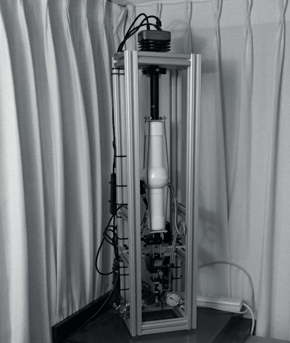
023. Multimodal anticipation and response to unpredictable car collisions in immersive virtual reality (ongoing)
A long-standing and debated question in this field is whether the body and brain can register a randomly chosen, emotionally important event a moment before it happens. Studies report such anticipatory effects, but they tend to be small and inconsistent from one replication to the next. One possible reason is that conventional experiments are highly sterile: participants sit still and view hundreds of flat images on a screen, conditions that bear little resemblance to how anticipation might operate in real life. We asked whether moving this question into a richer, more naturalistic setting, while tightening the experimental controls, would help. The study was preregistered at the outset.
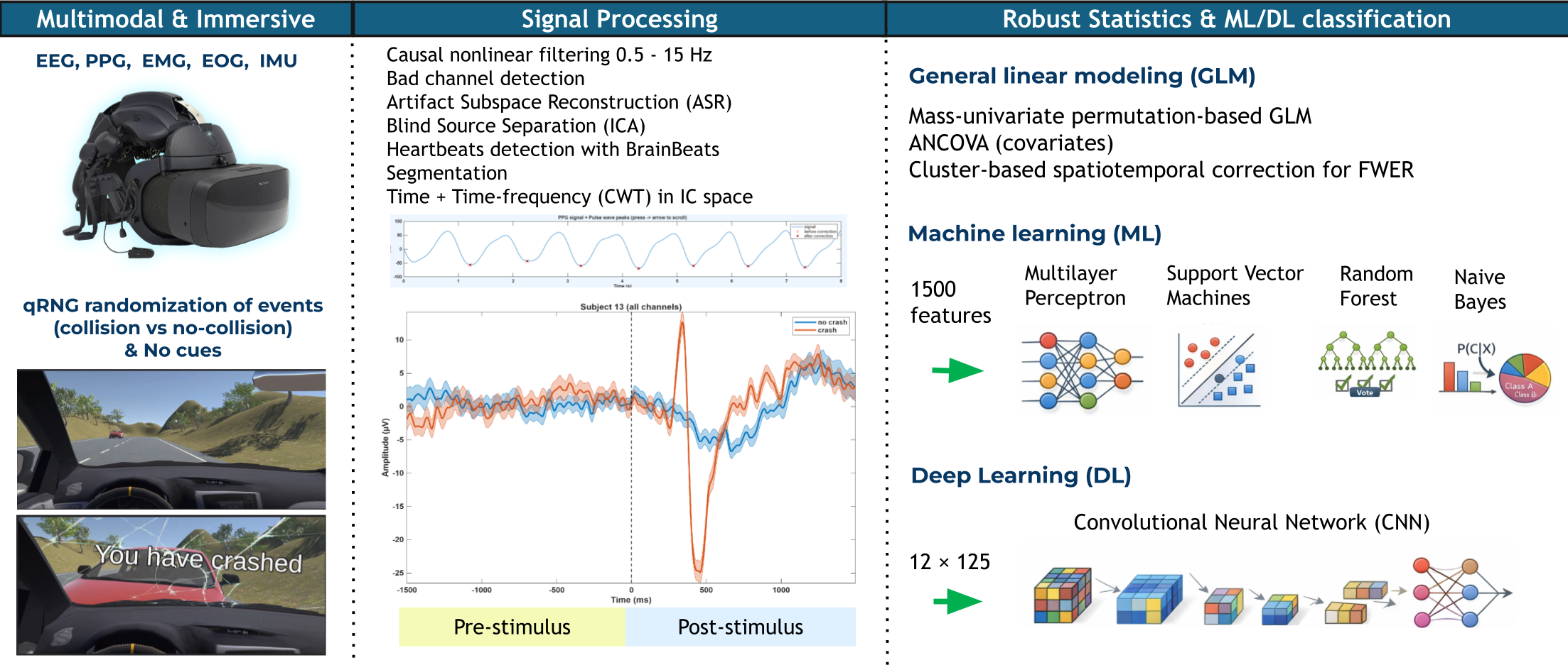
Working with the Galea headset (OpenBCI, Inc.), we built a fully immersive virtual-reality driving study that records several body signals at once: brain activity (EEG), heartbeats (PPG), eye movements (EOG), muscle activity (EMG), and head and body motion (IMU). Participants passively experienced driving trials with a genuine 50% chance of a collision (set by a quantum random number generator so that no pattern could be learned or expected in advance).
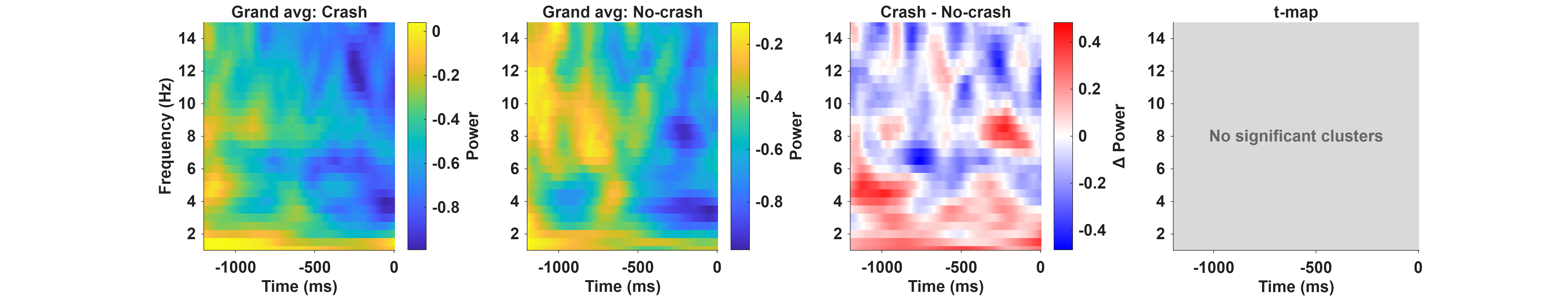

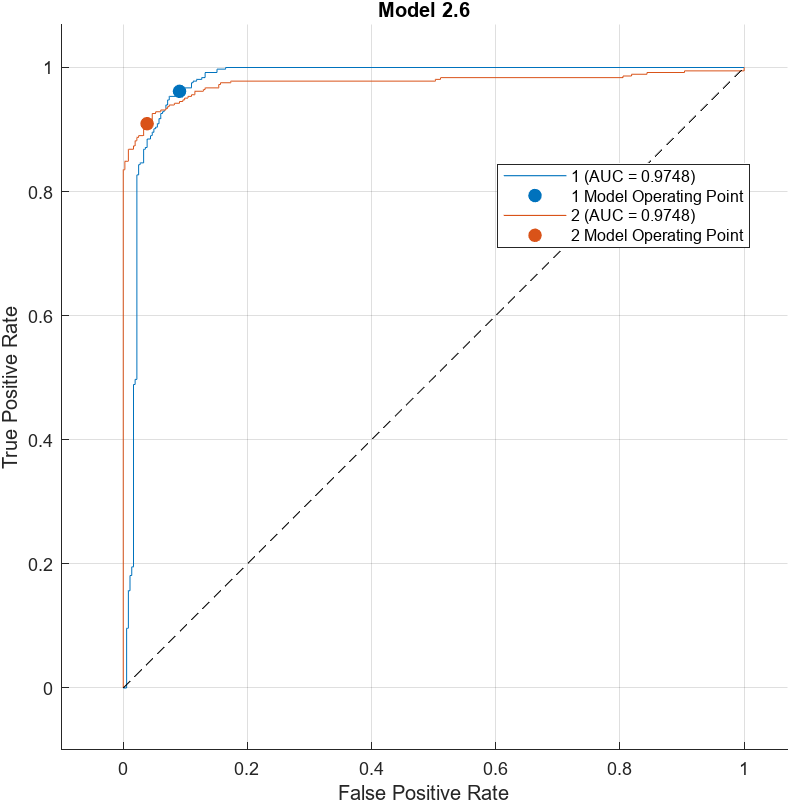
We found no statistically significant difference in the pre-stimulus (anticipatory) window once rigorous statistical corrections were applied. But unfortunately, the study had to be stopped mid-way due to hardware issues (mainly EDA), so the original sample size planned to detect such an effect was no longer met (N = 17 instead of 100). The clearest contribution of this work is therefore methodological: it shows that combining a novel multimodal wearable, immersive VR, and tightly controlled, bias-resistant design is feasible in practice, and it validates the recording and analysis pipeline through a strong post-stimulus result, where brain responses 238 to 754 ms after a crash were distinguished from non-crashes with up to 90% accuracy (shown in the figure). This establishes a robust template for future, adequately powered studies of anticipation in naturalistic settings. The full paper (Cannard & Yesilbas) is in preparation for peer review.
010. Brain predictive processes of emotional stimuli under unpreditable settings (ongoing)
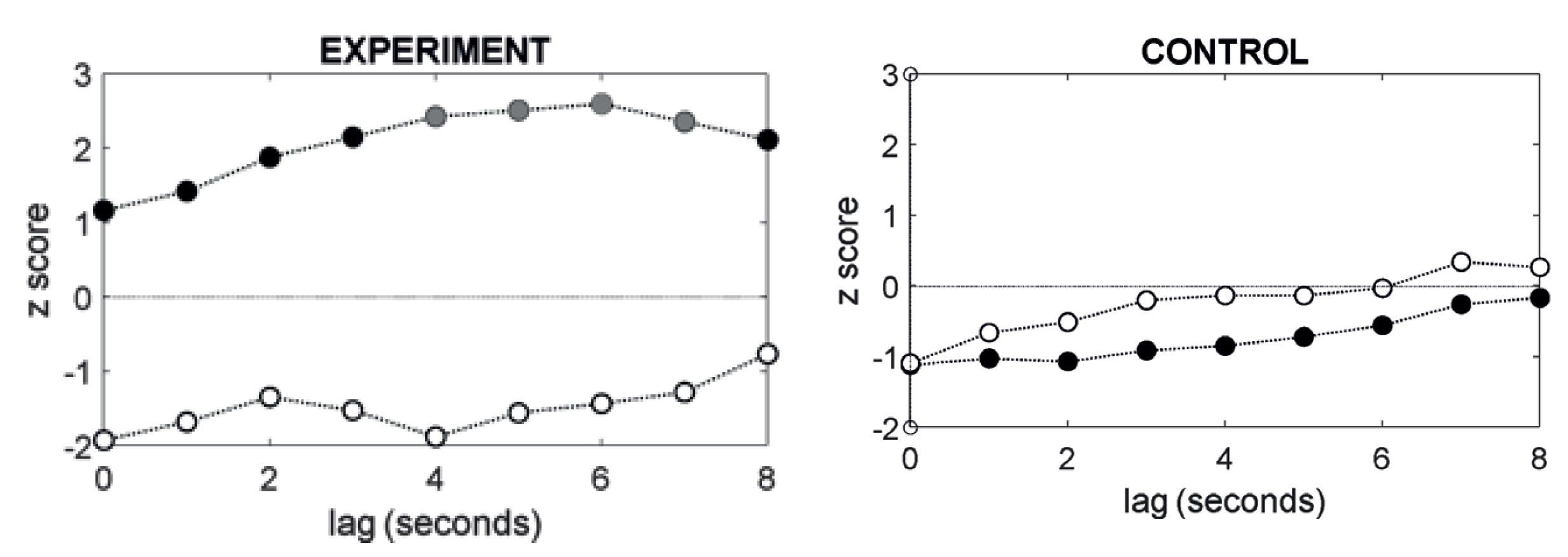
Can the brain prepare for something emotional before it even appears, even when there is no way to predict exactly when it will happen? The brain is known to anticipate events when their timing is regular, but far less is known about what it does in genuinely unpredictable settings. Using high-density EEG (64 channels, 81 participants) and rigorous, state-of-the-art statistical methods, we examined brain activity in the moments just before pleasant, unpleasant, or neutral images were shown.
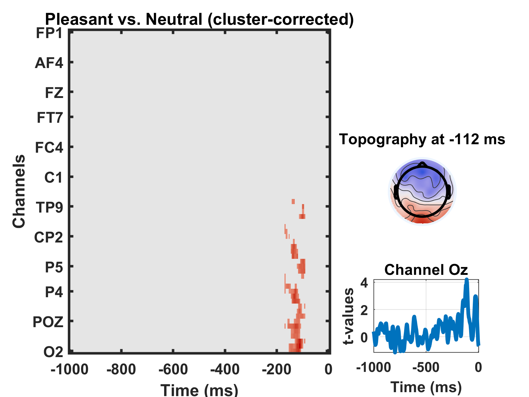
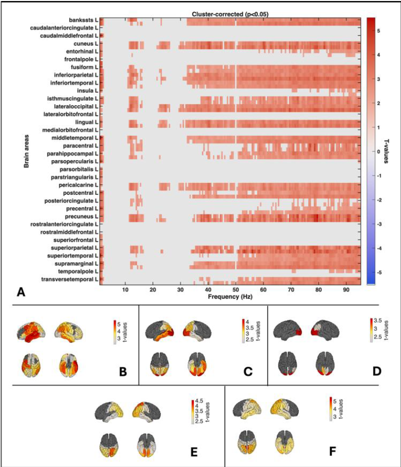
We found a clear anticipatory signal preceding pleasant images: roughly 170 to 90 milliseconds before a pleasant image appeared, activity over the right rear of the head (the occipital region) differed significantly from the activity preceding neutral images, peaking about 116 ms before onset. Source localization traced this signal to the right lateral occipital cortex, a region central to visual perception (shape, color, motion), object recognition, spatial processing, visual memory, and visual attention. In other words, the brain's visual system appears to "lean in" and prepare just before something rewarding is about to be seen, even without reliable timing cues.
Against our expectations, no equivalent anticipatory difference appeared before unpleasant images relative to neutral ones. We are now running follow-up analyses to rule out more ordinary explanations, such as effects of time-on-task, the order in which images were shown, or the brain implicitly learning the intervals between stimuli (quietly estimating, from past timing, when the next image is likely to occur).
014. Measuring brain entropy (ongoing)
Cedric developed an EEGLAB plugin to compute entropy measures at various time scales from biosignals (e.g., EEG, MEG, ECG, PPG). Such nonlinear measures are particularly promising for capturing complex multidimensional dynamics that may be missed by conventional linear measures (time or frequency analysis). Since these entropy measures are very computation-heavy, the plugin supports GPU and parallel computing when possible.
The plugin is for both novices (graphical user interface) and expert programmers and provides meaningful visualizations to ease interpretations of the results.
022. The OMNI image dataset (2026)
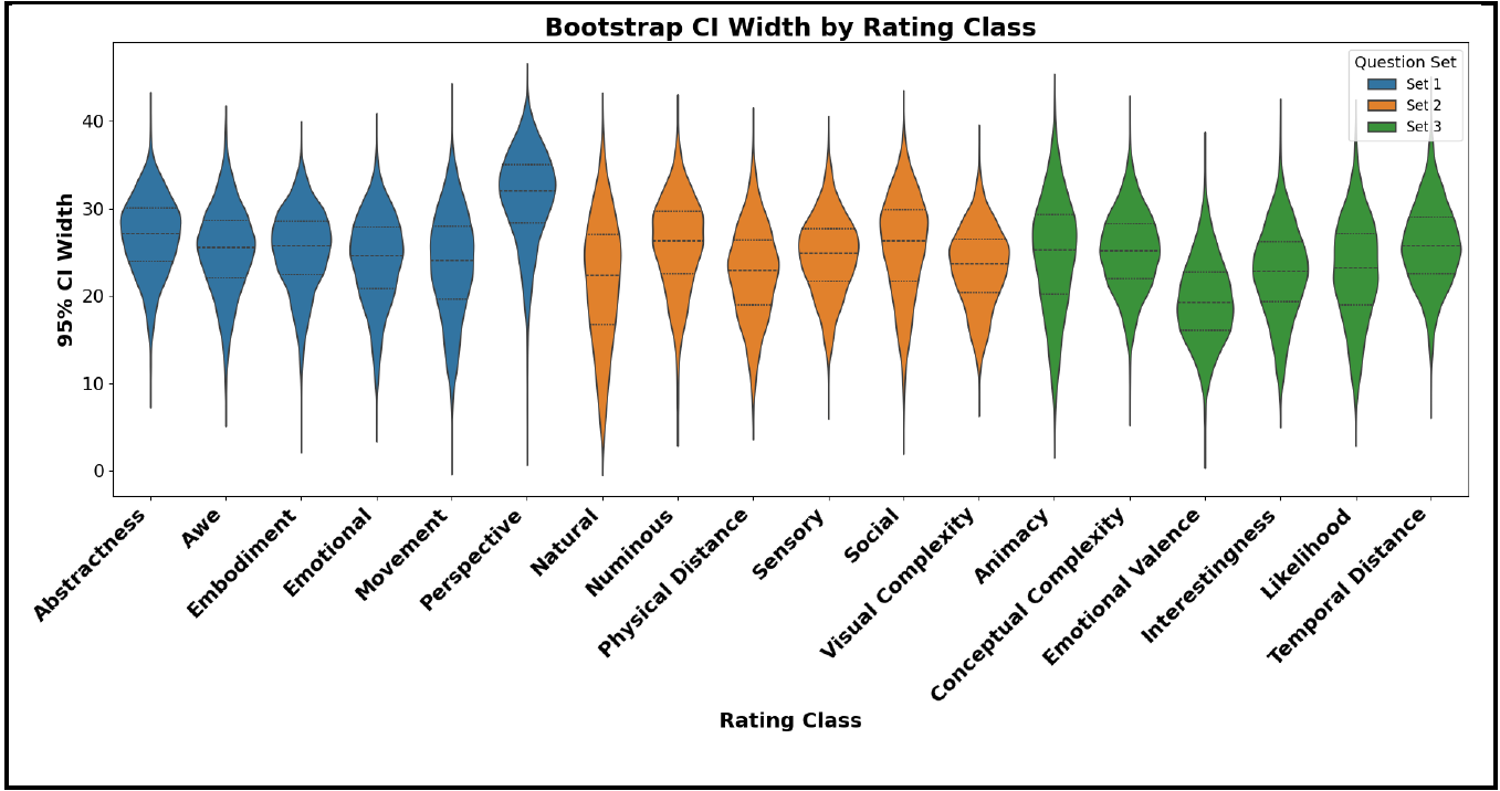
Researchers who study emotion, attention, or perception need standardized images whose properties are known in advance. We built the Open Multidimensional Normed Images (OMNI) dataset, a public repository of images rated by people on a wide range of cognitive, affective, and perceptual dimensions (for example emotional valence, arousal, visual complexity, and how interesting or socially relevant an image feels). These human ratings let other scientists select images with precisely the characteristics their study requires, making experiments easier to design and to reproduce. The dataset and ratings are openly available here (preprint currently under peer review).
020. Brain and body dynamics of the Wim Hof method (2026)
The Wim Hof Method combines bouts of fast, deep breathing with breath holds and cold exposure, and many practitioners report effects on stress and alertness. We recorded 64-channel EEG together with breathing, heart rate, and blood-oxygen measures in 17 adults (10 trained experts and 7 newcomers) during a standardized breathing session, and in the experts during brief cold-water immersion. The breathing lowered carbon dioxide in the blood (hypocapnia), and trained experts showed a stronger drop along with distinct brain-activity patterns, including increased gamma-band activity at the back of the head and reorganized communication between brain networks. The results suggest that practice changes how the breathing method engages both the autonomic nervous system and large-scale brain dynamics. Read the full article here.
021. Genomic correlates of self-reported extraordinary experiences (2026)
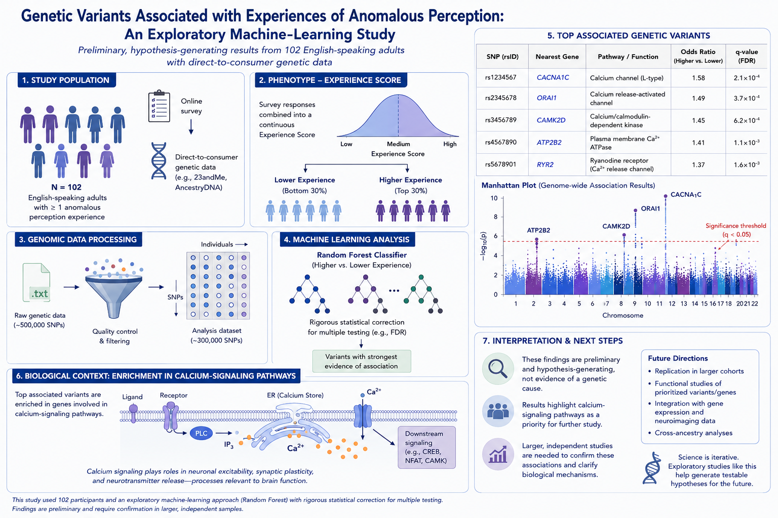
Extraordinary experiences are reported across cultures and often seem to run in families, which raises the question of whether biology plays any role. In this exploratory study, 102 English-speaking adults who reported at least one such extraordinary experience and had direct-to-consumer genetic data completed surveys and shared their genetic files. Using a machine-learning approach (a Random Forest classifier) with rigorous statistical correction, we looked for genetic variants (single-nucleotide polymorphisms, or SNPs) that distinguished people with lower versus higher experience scores. The analysis flagged a set of variants, several linked to calcium-signaling pathways, as worth investigating further. These are preliminary, hypothesis-generating results rather than proof of a genetic cause, and they point to directions for larger confirmatory studies. Read the full article here.
019. Lucid dreaming workshops for trauma and well-being (2025)
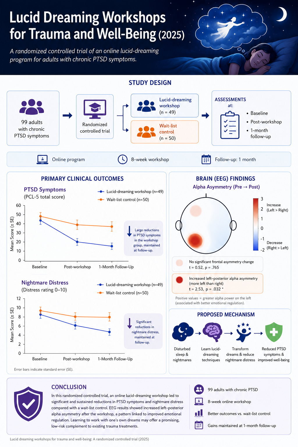
Lucid dreaming is a state in which a sleeping person becomes aware they are dreaming and can deliberately act within the dream. This line of work tested whether an online workshop teaching lucid-dreaming techniques could help people transform their nightmares and reduce trauma symptoms.
In a randomized controlled study, 99 adults with chronic PTSD symptoms were assigned either to the workshop (49 participants) or to a wait-list control group (50 participants). Compared with controls, the workshop group showed significant reductions in PTSD symptoms and nightmare distress, along with improved well-being, and these gains were maintained at the one-month follow-up. The findings suggest that learning to work with one's own dreams may offer a promising, low-risk complement to existing trauma treatments.
A companion EEG and HRV pilot study looked at what happens in the brain and body during these workshops, recording electrical brain activity (EEG) and heart-rate variability (HRV) to begin mapping the physiological changes that accompany lucid-dreaming practice.
018. The BrainBeats toolbox (2024)
I developed the BrainBeats toolbox, implemented as an open-source EEGLAB plugin to facilitate the study of brain-heart interplay, using EEG and cardiovascular signals (ECG/PPG).
Four methods are available: 1) heartbeats-evoked potentials (HEP) and oscillations (HEO), which captures how the brain responds to heartbeats with millisecond accuracy; 2) extraction of EEG and HRV metrics (time, frequency, and nonlinear domains) to find associations or differences between these features, or to assess Pre/Post-intervention changes; 3) Extract cardiac field artifacts (CFAs) from EEG signals with independent component analysis (ICA); and 4) calculate brain-heart coherence and causal interactions to better understand the interplay between the two systems. The toolbox is for both novices (general user interface) and experts (command line) and comes with a step-by-step tutorial.
The toolbox is open-source and available here
015. EEG and HRV correlates of well-being using low-cost wearable neurotech (2024)
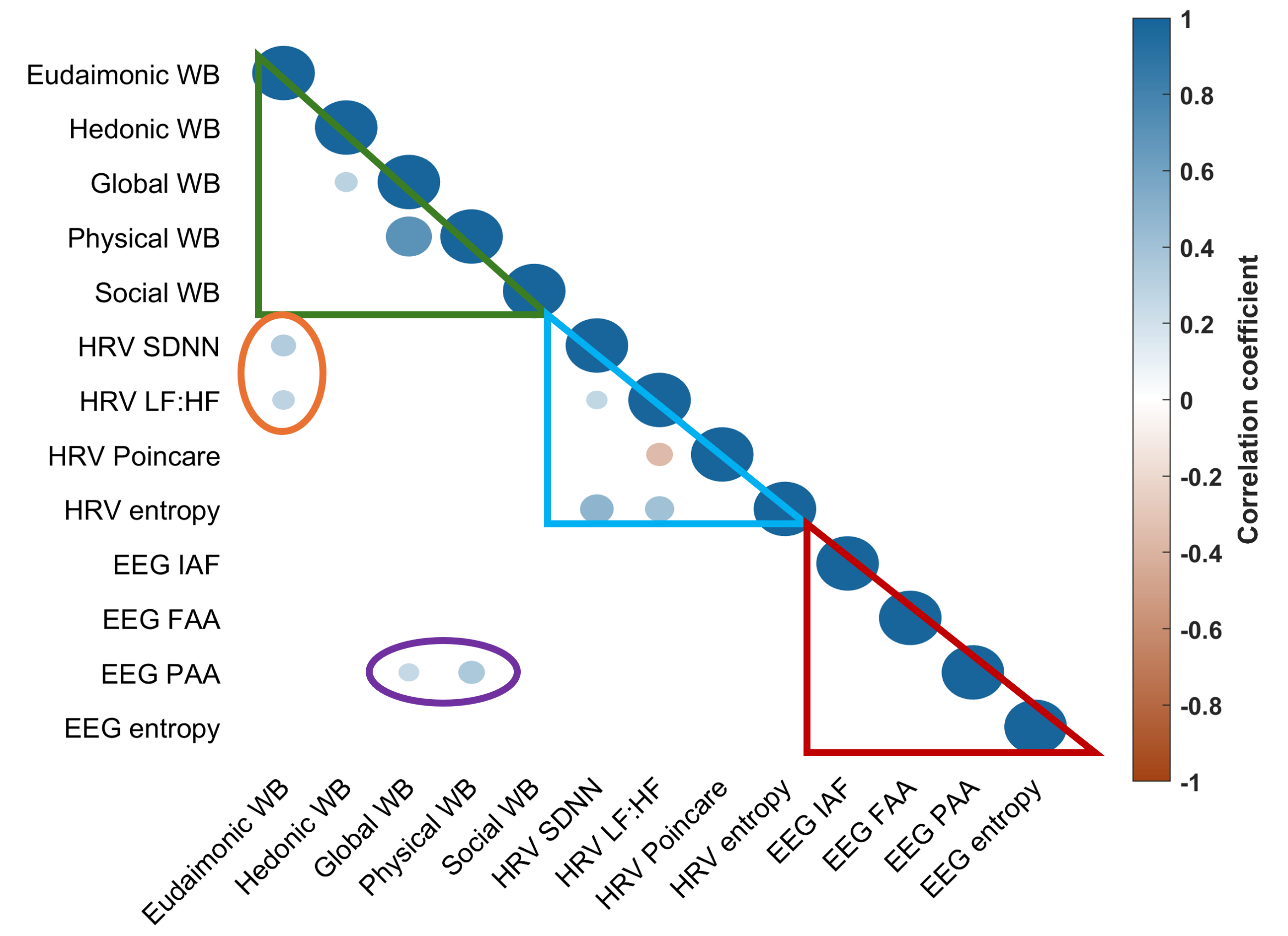
Wearable devices that measure brain waves (EEG) and heart activity (ECG) could be a simple and low-cost way to check how well people are feeling in everyday situations. However, it's tricky to get clear signals and find reliable indicators of well-being. We tested if these devices could identify markers of well-being by collecting short EEG and ECG recordings from 60 people in real-world settings. We looked for patterns in the data that might relate to different aspects of well-being, like happiness, life satisfaction, physical health, and social connections. Our study found some links between heart activity and psychological well-being, and between brain activity and physical well-being. These findings suggest that wearable devices could offer a quick and easy way to monitor well-being in daily life.

017. Scoring human creativity with AI (2024)
In this study, we evaluated the performance of an AI algorithm, the Open Creativity Scoring with Artificial Intelligence (OCSAI) system at scoring the Alternate Use Task (AUT), which tests some aspects of human creativity. We saw a strong correlation between manual (3 human raters per rating) and OCSAI scores for elaboration (rho = 0.76, p < 0.001, n = 520), indicating that the AI-based system effectively captures the elaboration component of creativity (i.e., the level of detail and development in responses). The correlation for originality component was weaker but significant (rho = 0.21, p < 0.001, n = 520), with originality referring to the uniqueness and novelty of ideas. These findings highlight the potential of AI-driven methods like OCSAI to automate the scoring for this task, especially for elaboration, while suggesting that further refinement may be needed for accurately assessing originality. Read the full article here.
016. Investigating an unusual visual experience (2024)
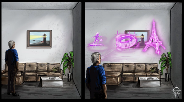
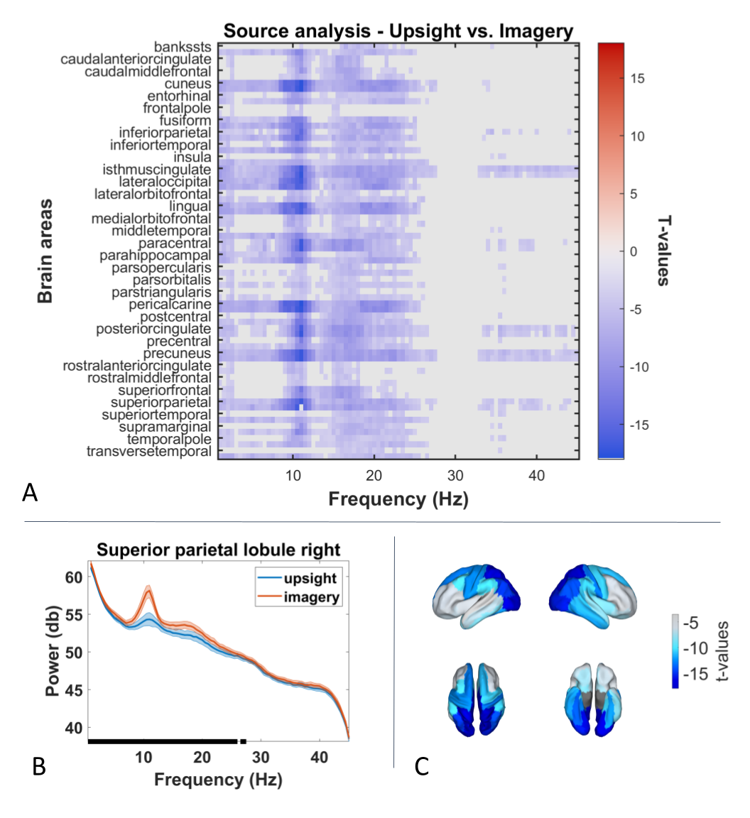
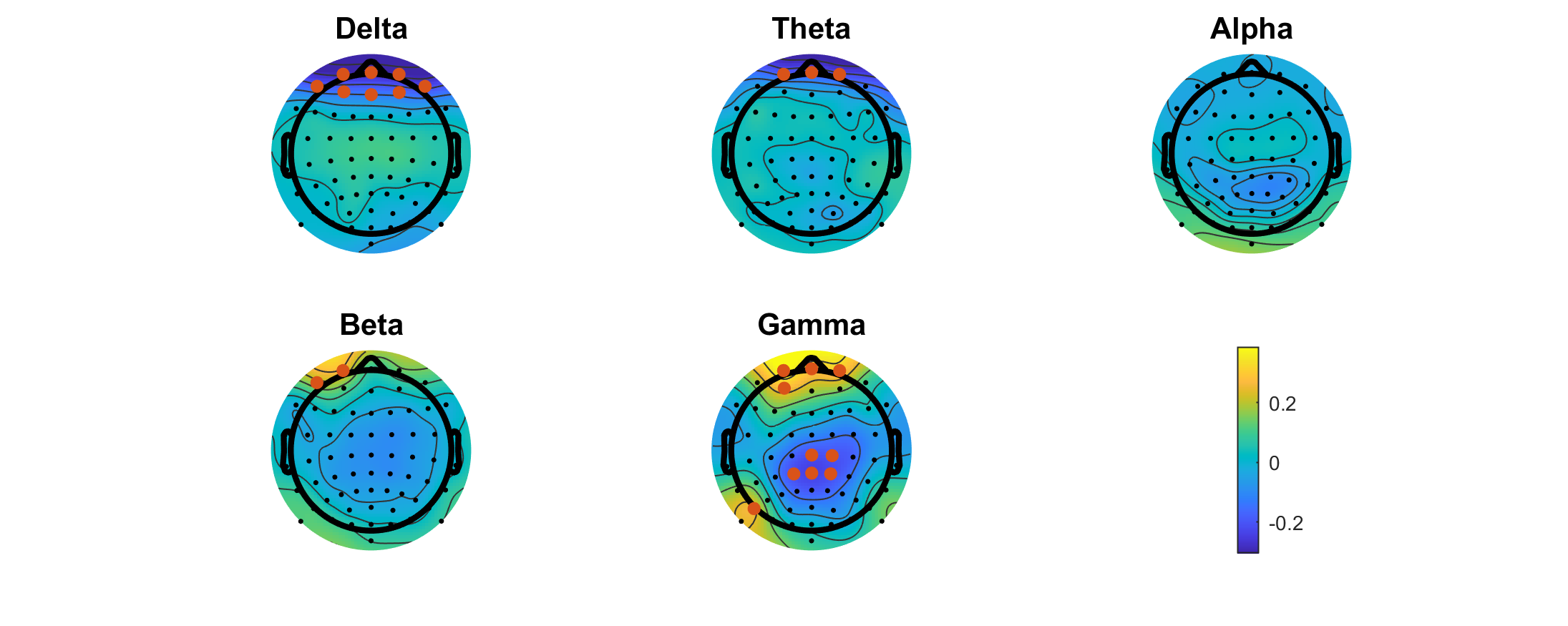
This case study explored a rare visual phenomenon where an individual continuously perceives vivid holographic images overlaid on their visual field and can partly control them. Using 64-channel EEG, we compared this experience with visual mental imagery across 200 trials. This experience showed marked decreases in alpha and delta power and increased gamma activity, alongside reduced alpha power and functional connectivity in visual and spatial processing neural networks (after source reconstruction to identify which brain areas were activated and interacting). These patterns suggest that this experience engages visual and cognitive systems more strongly than typical visual imagery while also differing from both imagination and visual hallucinations. Read the full article here.
013. Classifying EEG signals with machine learning (2023)
This project aimed to train a machine learning (ML) model to classify EEG signals (good/bad) collected with the MUSE headsets. First, I manually labeled 3000 30-second segments and extracted features in the time, frequency, and nonlinear domains. Then I trained an ensemble of ML models (decision trees, logistic regression, LDA, SVM, Naive Bayes, neural networks) implementing feature-selection, PCA-dimension reduction, hyperparameter tuning, and 5-fold cross-validation (on 80% of the data). The best model was then validated on a test dataset (20% remaining data from different subjects to avoid overfitting). The best models achieved 93.5% of accuracy for frontal channels and 91.4% for the posterior channels. These classifiers can be used via my import_muse plugin to classify bad MUSE channels during importation.
012. Advanced re-referencing methods for EEG data (2021)
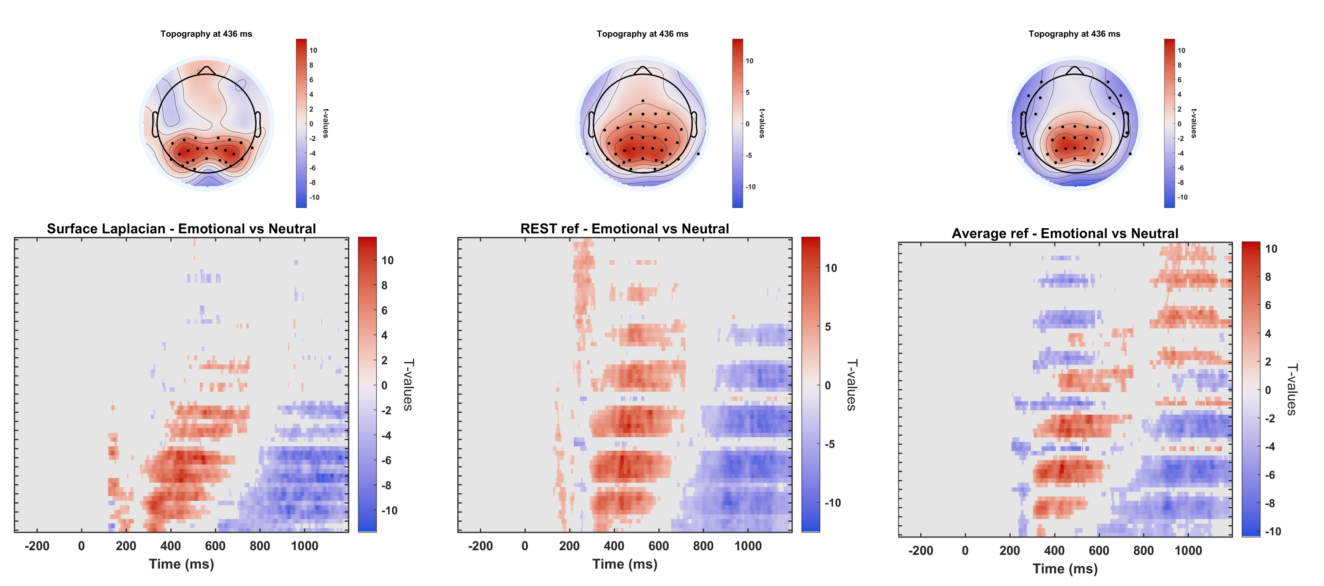
I implemented two advanced re-referencing methods for multidimensional EEG data: the reference electrode standardization technique (REST; Yao 2001) and the Surface Laplacian transformation (also called current-source density transformation). The image illustrates the statistically significant differences at the grand average ERP level (N = 81, 230 trials each, after spatiotemporal cluster correction, p = 0.01), comparing the brain's response to emotional stimuli (pleasant and unpleasant valence with high arousal) relative to neutral ones (no emotional valence and low arousal). We can see that the reference to infinity (REST) is preferable to the commonly used reference to average when assessing global (widespread) scalp dynamics, whereas the Surface Laplacian is best to capture local dynamics, dealing better with undesired volume conduction effects (especially for source analyses).
Both are accessible via command line for easy implementation and automation.
011. 3D ERP video (2021)
3D visualization of the spatial distribution of an averaged event-related potential (ERP) over several hundred milliseconds from -100 to 450 ms after stimulus presentation (at time = 0). Current-source density (CSD; i.e., Surface Laplacian) transformation was performed on this subject's 64-channel EEG data to increase spatial resolution and local dynamics.
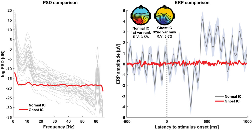
009. Avoiding EEG "Ghost ICs" (2023)
Many EEG researchers use independent component analysis (ICA) to process and analyze their EEG data. However, some common processing steps (e.g., electrode interpolation or re-reference to average) can lead to effective rank deficiency, which in turn, leads to the emergence of "ghost ICs". These independent components display a typical scalp topography, but actually contain white noise properties in both time and frequency domains. They can therefore be easily missed and significantly affect findings in unknown ways.
See the full-article here for more details.
008. Importing EEG data (2022)
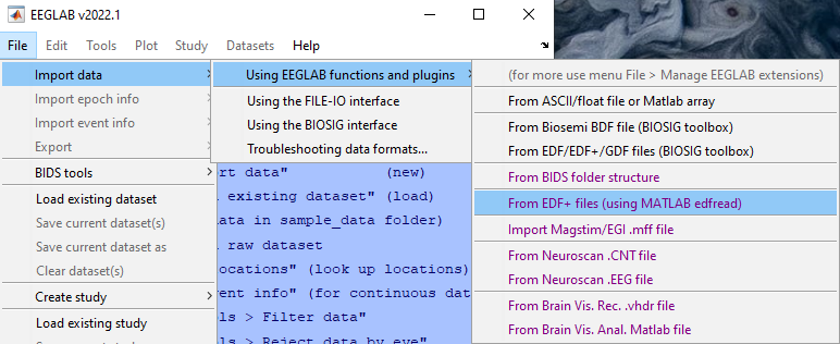
I developed the import_muse() and import_edf() tools to import data files from various formats (.csv, .edf, .edf+, etc.) into EEGLAB. Implemented as EEGLAB plugins, they can be used with the GUI or command line. The plugins detect the data types (e.g., EEG, PPG, GYR, ACC, AUX, etc.), the sample rate, annotations if any, etc. and convert everything into the EEGLAB format. Step-by-step tutorials are available.
006. Validating EEG signals from a wearable system (2021)
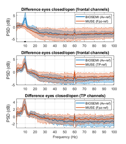
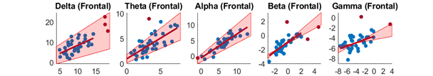
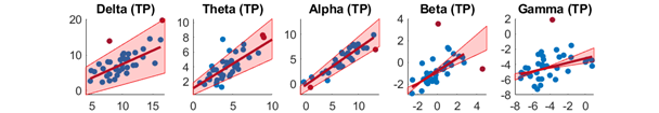
This project aimed to assess whether the Muse (InteraXon, Inc.), a wearable EEG headset, can be used to reliably capture EEG signals and relevant measures (alpha asymmetry and individual alpha frequency). A minimal amount of data was deliberately collected to test the feasibility for real-world applications (EEG setup and data collection being completed in under 5 min). The MUSE's temporoparietal (TP) channels showed comparable power spectra, alpha asymmetry, and individual alpha frequency (IAF) relative to that obtained from a gel-based state-of-the-art BIOSEMI system (referenced to both Fpz like the Muse, and to average, the gold standard). However, the frontal channels needed to be re-referenced to linked-mastoids to provide valid outputs. Furthermore, we observed satisfying internal consistency reliability. See the publication here for more details.
Mastoid-ref montage (frontal channels)
Fpz-ref montage (TP channels)
007. Consciousness, light, and the Observer Effect (2021)
The double-slit experiment in quantum mechanics demonstrates how observation influences particle behavior, suggesting that consciousness may play a role in collapsing the wave function. Light polarization, the orientation of light waves as they oscillate perpendicular to their direction of travel, is a key property of light. Studying these aspects of light—polarization, scattering, and intensity—provides insight into fundamental physical processes and the nature of observation itself. To test whether focused attention could alter light’s polarization, we passed a laser beam through two polarizers and asked participants to direct their attention toward and away from it. Instead of increasing intensity as expected, the light unexpectedly dimmed. To explore whether attention affects how light scatters or is absorbed—phenomena important in atmospheric optics and medical imaging—we then passed a laser beam through a reflective sphere and measured both transmitted and deflected light. Surprisingly, the beam’s intensity increased after exiting the sphere. These results parallel the observer effect in quantum mechanics, raising further questions about the connection between consciousness, perception, and physical reality.
See the full-article here for more details.
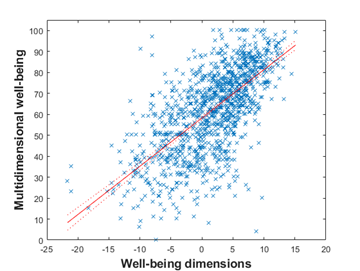
005. Identifying the main well-being dimensions (2021)
This project aimed to identify the main dimensions of multidimensional well-being (MWB). 2647 individuals participated in an online survey between November 19, 2020, and September 26, 2021, during the COVID-19 pandemic. 1615 were unique pre-intervention records and 429 were unique post-intervention. After validating a quick visual analog scale for capturing multidimensional well-being in a single question (convergent and test-retest validity), a multiple regression model showed that the main dimensions of MWB were the hedonic WB (i.e., affective WB), eudaimonic (psychological WB), physical WB (i.e., health and pain levels), and social (sense of connectedness with others) dimensions. The model explained 44.6% of the variance in MWB (N = 1443).
Multidimensional well-being was also influenced by reported connection with nature, religion/spirituality, physical activity during leisure, and personality trait (N = 415). Physical activity at work, meditation practice, relationship status, and creativity levels did not show any relationship with MWB.
See here for more details.
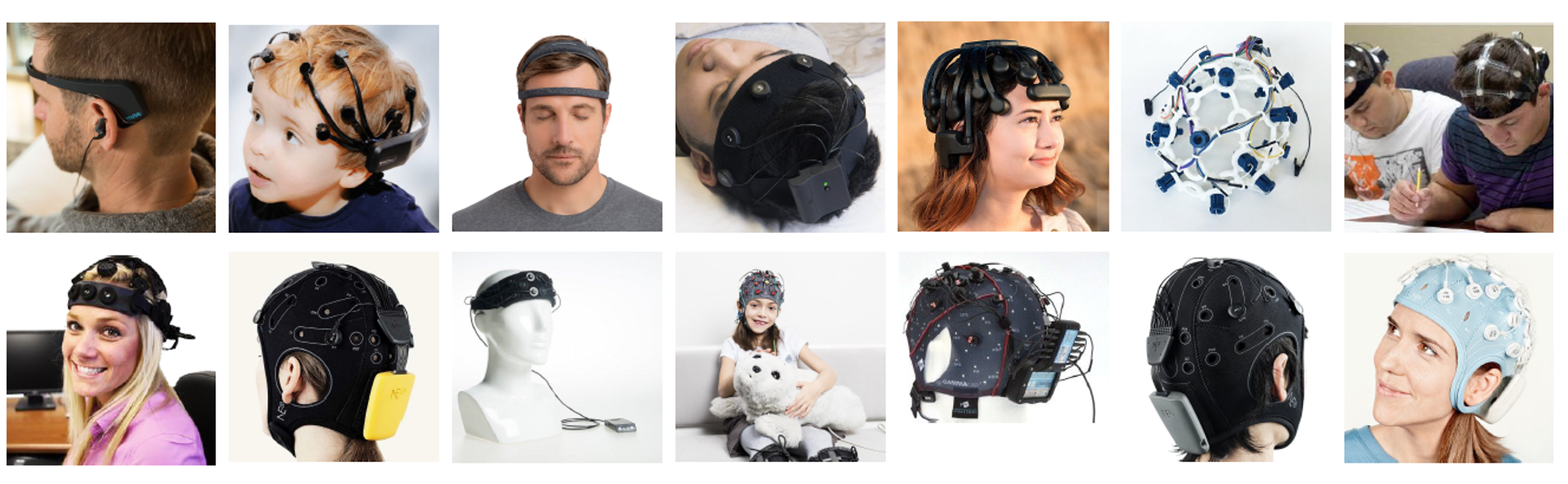
004. Self-health monitoring and wearable neurotechnology (2020)
BCIs and wearable neurotechnologies are being used to collect real-time neural and physiological data, showing great promise for advancing medical diagnostics, prevention, and intervention. This book chapter discussed here presents wearable electroencephalography systems, highlighting their groundbreaking innovations in real-time health monitoring and their technical pros and cons. Though these systems offer potential for large-scale data collection beyond traditional laboratory settings, they also face methodological and ethical challenges that must be addressed in the current and future research.
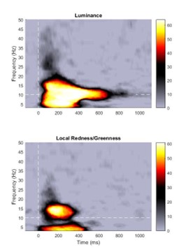
002. Brain oscillations in color vision (2016)
I did my 2nd Master's project with Dr. Rufin VanRullen. In a previous study, Rufin identified the brain's preferred response to luminance, the so-called "echo function" (an EEG oscillation displaying reverberations at 10 Hz up to 1 s after the presentation of the stimulus; see VanRullen & MacDonald (2012) ). The goal of my Master's project was to determine whether this echo functions was also the brain's preferred way of processing color stimuli (namely red, green, blue, yellow). The project involved developing psychophysics code to find individualized equiluminance threshold, modifying stimulus presentation scripts, and recording and analyzing 64-channel EEG on 12 subjects.
Results suggested that no equivalent echo function exists for color vision.
003. Physiological examination of an altered state of consciousness (2018 & 2023)
Some individuals are able to enter deep trance states at will, where their consciousness is highly altered (no awareness, no recollection, feeling loss of control over the body, etc.). We assessed how physiological activity (64-channel EEG, ECG, GSR, voice) would differ between this state and a baseline (directed mind-wandering).
A first analysis showed differences only in the voice data.
A second analysis addressed some limitations and showed increased EEG delta and theta power in the frontal region and increased gamma in the centro-parietal region during mind-wandering, whereas trance showed increased beta and gamma power in the frontal region. A source-level analysis (spectral power and functional connectivity across brain regions) showed no differences. However, reported trance depth was negatively correlated with whole-brain connectivity in all frequency bands, suggesting that deeper trance was associated with less overall brain functional connectivity.
001. Brain atrophy in Schizophrenia (2015)
During my first research project with Dr. Sonia Dollfus (1st year of MS.c.) at the ISTCT lab in Caen, France, I learned how to process and analyze MRI data to measure grey matter atrophy in patients with Schizophrenia.
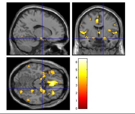
3T MRI scans were segmented and normalized using DARTEL in SPM12, allowing tailored template of the study population, reducing normalizing errors and enhancing alignment of small structures. After smoothing, intracranial volume was calculated using GM, WM, and CSF. A voxel-based morphometry (VBM) analysis (voxel-by-voxel comparisons) was conducted to characterize gray matter differences between the two groups (63 controls vs 51 patients), accounting for age, gender, education, and intracranial volume as covariables.
Whole-brain analysis showed atrophies consistent with the literature (medial orbitofrontal, bilateral insula, and left middle cingulate cortices; p-corrected = 0.001).
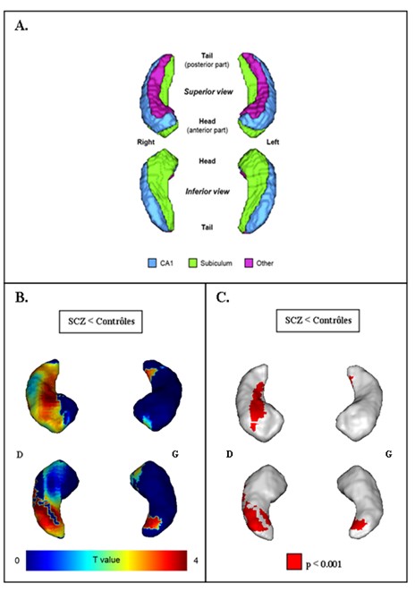
Then, the contrast map was projected onto a 3D mesh of the hippocampus and its subfields. This mesh was manually delineated on coronal slices by Renaud La Joie (see publication here ). In the right hippocampus, atrophy was detected in the median-posterior part of the Subiculum and other subfields, namely CA2-3 and DG-CA4, as seen in the dorsal view. This extended across almost the entirety of the CA1 region and marginally into the ventral Subiculum. In contrast, the left hippocampus showed a milder atrophy primarily concentrated in the posterior section of the Subiculum, discernible both ventrally and slightly dorsally. However, these differences did not survive the correction for family-wise error (FWE).